- Today
Hip Impingement (FAI): What It Actually Is — And Why the Diagnosis Gets Handed Out Too Often
- Kolten Tea
- Hip Labral Tears
- 0 comments
If you’ve been told you have hip impingement, or FAI, you’ve probably heard one of two versions of it. Either it’s a structural problem with the shape of your bones and you’ll eventually need surgery. Or it’s nothing to worry about, lots of people have it, go do some stretches.
Neither one is quite right, and the gap between them is where most of the confusion lives.
Here’s the thing that makes this diagnosis unusual: the shape of your hip and the pain in your hip are two separate facts, and they don’t automatically belong to each other. Plenty of people have the shape and no pain at all. Some people have the pain and the shape is incidental. Sorting out which one you are is the entire problem, and it’s not something a scan can do by itself.
So this article does four things. First, what FAI actually is — the three types, and why yours matters more than you’ve probably been told. Second, what it feels like, including the one finding that genuinely separates it from the other things that hurt in the same place. Third, why this diagnosis is handed out more often than it should be, which is the part I most want you to read. And fourth, what to actually do about it, starting today.
What FAI Actually Is
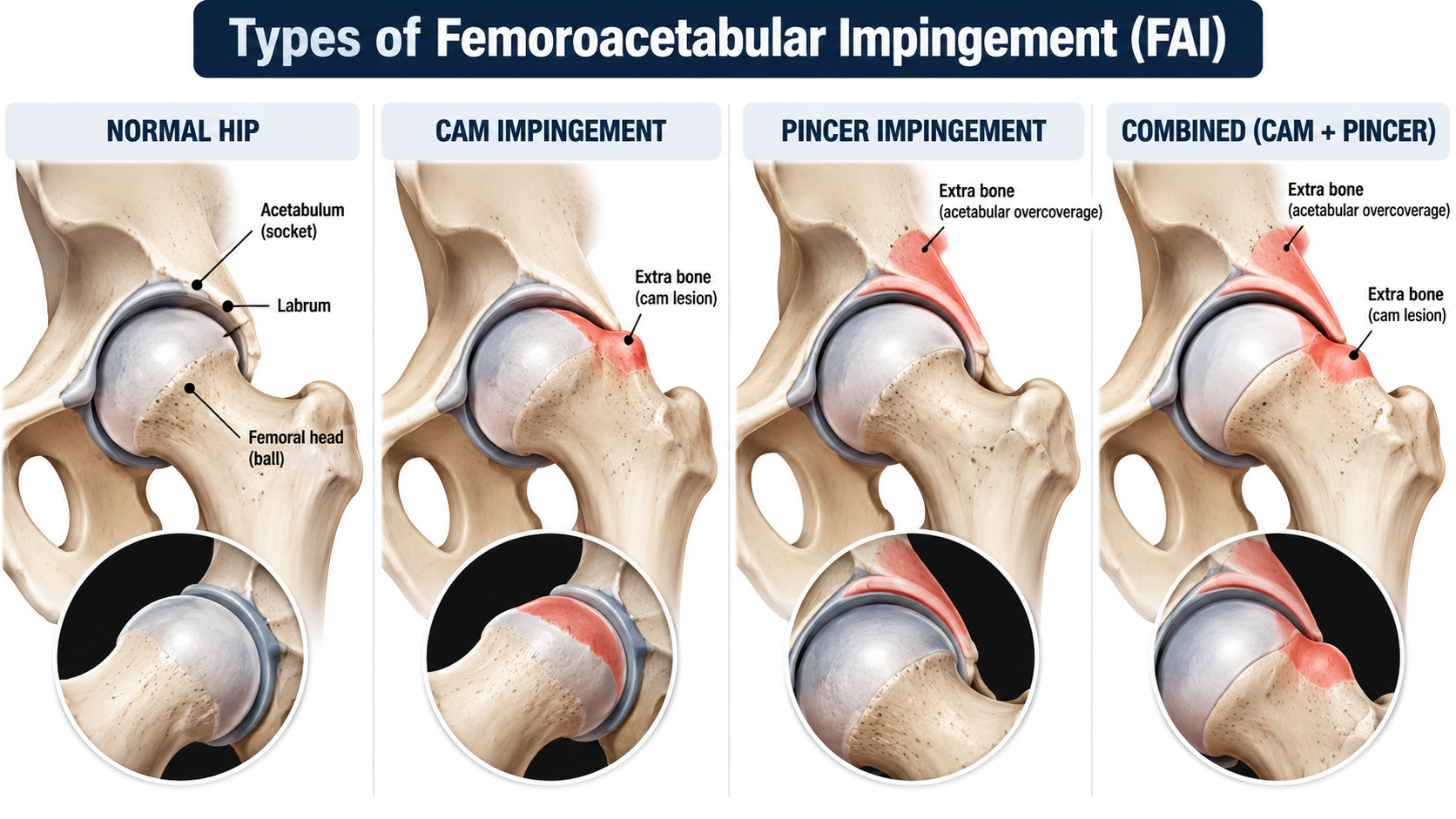
Your hip is a ball and socket joint. The ball is the top of your femur. The socket is part of your pelvis. In a hip shaped the way most textbooks draw it, that ball rotates smoothly inside the socket through a full range of motion without anything bumping into anything.
Femoroacetabular impingement is what happens when the shapes don’t allow that. There’s premature contact between the ball and the rim of the socket before you reach the end of the motion. The joint runs out of room early.
There are three ways that happens.
Cam impingement
The ball isn’t perfectly round. There’s extra bone along the junction where the head of the femur meets the neck, so instead of a sphere rotating cleanly inside the socket, you have a slightly oval shape that jams into the rim when you flex and rotate.
Cam is more common in young athletic men, and it’s associated with high activity during the years the skeleton is still developing.
Pincer impingement
Here the ball is fine — the socket covers too much of it. Either there’s extra rim, or the socket is oriented in a way that overhangs the ball. Instead of the ball jamming into the rim, the rim is already sitting in the way.
Pincer skews more common in women, and often slightly older.
Mixed
Both at once. And this isn’t some exotic third category — mixed is common. A lot of people have some of each.
Why the type matters
This is the part that usually gets skipped, and it’s the reason the distinction is worth making at all.
The two types do different damage.
Cam tends to shear the labrum and the cartilage underneath it, from the inside out. The oval bump sweeps past the rim and peels the tissue off the bone.
Pincer tends to crush the labrum between two bones. The rim is in the way, and the labrum gets pinched against it.
Same diagnosis on paper. Different injury pattern underneath. Which means a different conversation about what to expect and what to do.
Where the extra bone comes from
This is the question I get immediately after explaining all of the above: if I never injured my hip, where did the extra bone come from?
Short answer — you grew it.
Cam morphology develops during adolescence, while the growth plate at the top of the femur is still open and still responsive to load. It’s an adaptation to what you were doing with your body during those years, not an injury that happened to you. That reframe matters, because people hear “extra bone” and assume something went wrong. Mostly, something adapted.
What It Actually Feels Like
If you’ve read my article on labral tear symptoms, some of this is going to sound familiar, and there’s a good reason for that. FAI is one of the things that causes labral tears in the first place. The labrum gets torn by the abnormal contact, so the symptoms overlap heavily.
Where the pain shows up
Deep groin pain is the most common presentation, by a wide margin. People usually describe it as mechanical — tied to specific positions and movements rather than aching constantly in the background.
It can also refer into the thigh, the back, or the buttock.
Beyond the pain itself, the symptoms people commonly report are clicking, catching, stiffness, a sense of buckling or giving way, and limited range of motion.
What makes it worse
The provoking positions are the ones that close down the front of the joint:
Deep squatting
Prolonged sitting, especially in low seats
Getting in and out of a car — the one almost everybody recognizes
Pulling your knee toward your chest
Pivoting or cutting in sport
The one finding that separates FAI
Here’s what distinguishes this from most of the other things that cause pain in the same neighborhood: limited range of motion. Specifically, limited internal rotation of the hip when it’s flexed.
Think about why that’s informative. A muscle strain hurts, but it doesn’t change how far your hip can physically rotate. A bone bumping into another bone does. That’s a mechanical block, and it’s one of the more telling things I look for in the clinic.
If your hip hurts but rotates freely, that points somewhere other than impingement.
Why This Diagnosis Gets Handed Out Too Often
Now the part I really want you to hear.
FAI has become one of the most over-applied diagnoses in hip care. Not because clinicians are careless — because there’s a genuine problem with what imaging shows.
Most people without hip pain have these findings
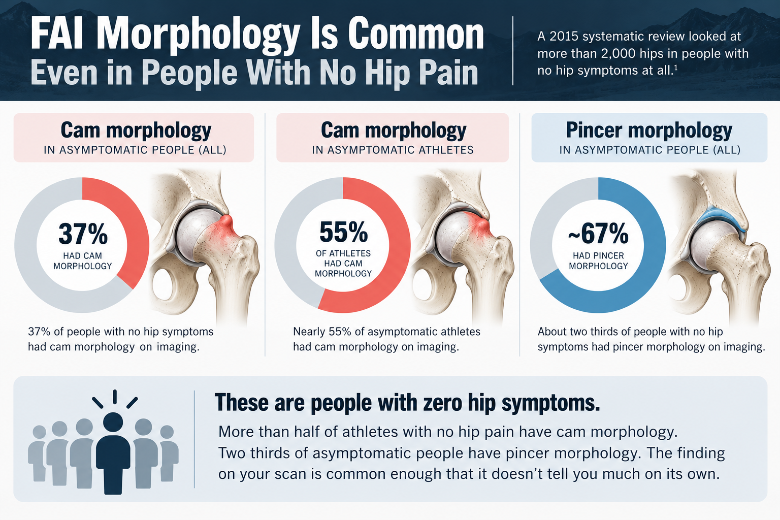
A 2015 systematic review looked at more than two thousand hips in people with no hip symptoms at all.1
Cam morphology showed up in 37% of them overall. In athletes specifically, nearly 55%. Pincer morphology showed up in about two thirds.
Read those numbers again. More than half of athletes with no hip pain whatsoever have cam morphology on imaging. Two thirds of asymptomatic people have pincer morphology. These are people with zero symptoms who were scanned anyway for research purposes.
So the finding on your scan is common. Common enough that finding it doesn’t tell you much on its own.
The alpha angle, and why the cutoff keeps moving
It gets more interesting when you look at how a cam is actually measured.
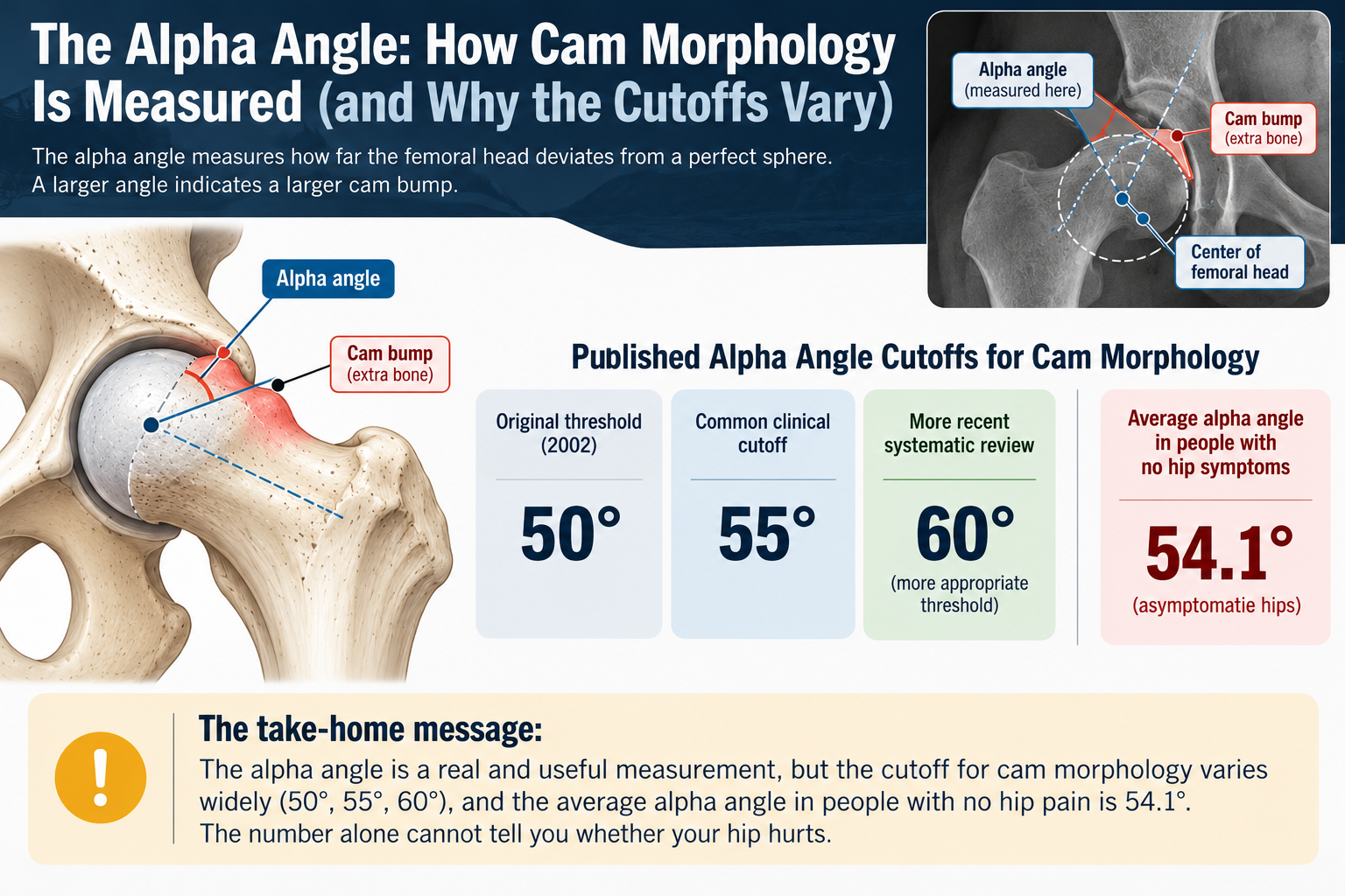
The measurement is called the alpha angle — essentially, how far the bump deviates from a perfect sphere. Bigger number, bigger cam.
Here’s the problem. The field doesn’t agree on where the cutoff should be.
The original threshold, proposed in 2002, was 50 degrees. The commonly used clinical cutoff drifted to 55. A more recent systematic review concluded that 60 is the more appropriate number.2
Now put that next to this: in that same review of asymptomatic hips, the average alpha angle was 54.1 degrees.1
That average — in people with no symptoms at all — sits essentially right on top of the common clinical cutoff.
That’s why the thresholds keep getting revised upward. The measurement is real and it’s useful. But the number alone cannot tell you whether your hip hurts.
So if you got imaging for hip pain and the report came back saying cam morphology, that finding might be causing your pain. Or it might be a feature of your anatomy that’s been sitting there for fifteen years and has nothing to do with why you hurt right now.
What a real FAI diagnosis requires
This is exactly why the field changed its language.
In 2016, an international consensus group published what’s called the Warwick Agreement, endorsed by 25 clinical societies.3 They introduced the term FAI syndrome specifically to put the patient’s symptoms back at the center of the diagnosis.
Under that agreement, a diagnosis requires all three of the following:
Appropriate symptoms
Positive clinical signs on examination
Imaging findings
All three. Not imaging alone.
The shape of your hip, by itself, is a finding — not a diagnosis. If you have cam morphology on a scan but no symptoms and no clinical signs, you don’t have FAI syndrome. You have a hip shape, and so does roughly a quarter of the general population.
What this means practically: if someone showed you a scan and said “there’s your problem” without examining you and without asking detailed questions about what provokes your pain, that’s an incomplete diagnosis. You’re allowed to ask about the other two pieces.
What to Actually Do
Let me be straight with you about what the research says here, including the parts that don’t flatter my own profession.
The honest part about surgery
There have been real randomized trials comparing hip arthroscopy to physiotherapy-led care for FAI syndrome. The largest one, run in the UK, found that at twelve months the surgical group scored better on hip-related quality of life than the group that received best conservative care — and the difference was large enough to be clinically meaningful.4
So I’m not going to tell you surgery doesn’t work. It does, for the right person.
But two things sit alongside that result.
Both groups improved. Conservative care wasn’t a failure in that trial. It was less effective on average, which is a different claim.
Surgery carries cost, risk, and a recovery measured in months. That’s a real tradeoff when the alternative also works for a lot of people.
The question to ask your surgeon
If surgery is on the table, there’s one piece of evidence that should change what you ask about.
The leading cause of revision hip arthroscopy — needing a second operation — is residual cam left behind from an incomplete resection the first time. In one study of fifty revision cases, 90% had leftover deformity.5
Think about what that tells you. The thing that predicts a good surgical result isn’t how big your cam was going in. It’s whether enough of it came out.
Which means the useful question for a surgeon isn’t “is my cam bad enough for surgery.” It’s:
“How do you confirm you’ve resected enough before you finish?”
That’s a question a good surgeon will have a real answer to.
Two principles before the exercises
For most people, unless there’s a clear reason otherwise, the reasonable starting point is loading the hip properly first. Two ideas drive everything below.
Work the hip in ranges that don’t jam it. This is the piece most people get backward. If your hip impinges in deep flexion with rotation, then grinding into deep squats and aggressive knee-to-chest stretching is loading the exact position that hurts you. Train strength in the ranges you have, not the ranges you’re trying to force.
Build the glutes, especially the abductors and deep rotators. These control how the femoral head sits and moves inside the socket. Better control means less of the abnormal contact causing the problem in the first place.
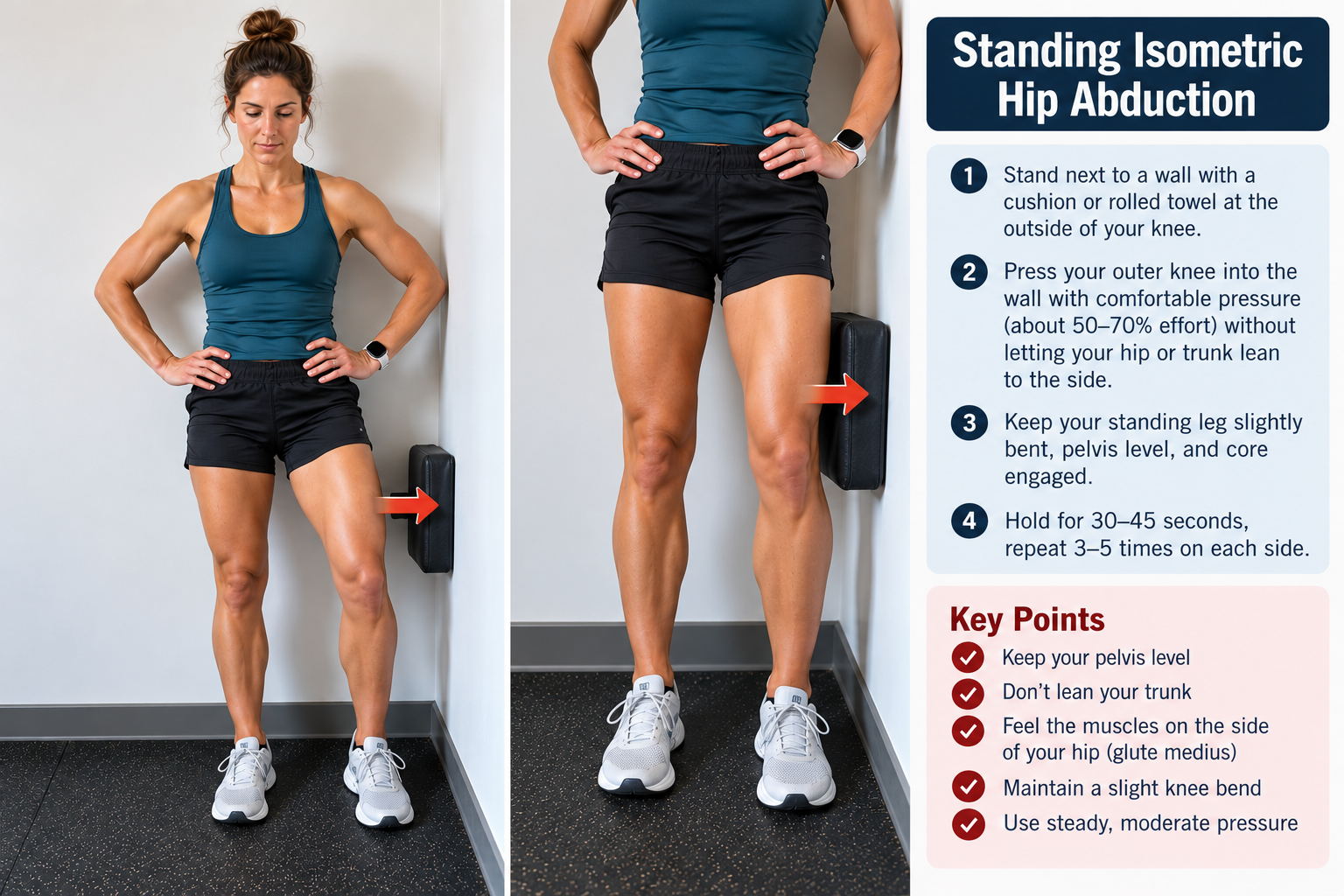
1. Isometric hip abduction (wall press)
Stand next to a wall. Bend your inside knee to about 90 degrees and press it into the wall.
30-second holds, 5 rounds each side.
You should feel this in the outside of your standing hip — the one on the floor, not the one pressing.
2. Single-leg bridge
On your back, one foot planted, the other leg lifted. Drive through the planted heel and lift your hips, keeping them level. Squeeze at the top.
10–15 reps, 3 sets each side.
If your hips tilt as you lift, drop back to a double-leg bridge until you can hold them square.

3. Controlled reverse step-down
Stand on a step. Slowly lower the opposite foot toward the floor behind you, taking 5 to 10 seconds on the way down, then come back up.
8–12 reps, 3 sets each side.
Slow is the whole exercise. If you’re dropping fast, lower the step.

The rules
Four to five days a week.
Be cautious with deep hip flexion for now. Not forever — for now.
Use the next-day rule. Mild soreness that settles within 24 hours is fine and expected. Pain that’s worse the next morning means reduce the volume.
Give it a real 6 to 8 weeks before judging it. Hip rehab for impingement runs on a longer timeline than most people expect, and most people quit well before the point where it would have started working.
And if you do all of that consistently and you’re still limited, that is genuinely useful information for a conversation about surgery. Not a failure. Information.
One Last Thing
This article can’t diagnose you. I haven’t examined you, I haven’t watched you move, and I haven’t seen your imaging. What it can do is help you understand what the finding on your scan does and doesn’t mean, and give you better questions to ask the person who has examined you.
If your pain is severe, getting worse, or started with a significant injury, get evaluated in person.
Want the full program? The Hip Labral Recovery System walks through the entire progression — what to do in each phase, when to advance, when to hold, and what to avoid.
Want someone to look at your specific situation? I offer one-on-one virtual consultations where we review your imaging, your history, and what’s actually provoking your pain—including whether what you’ve been told about that scan holds up.
Related reading
Get free PT tips before you need them. Once a month, I send out what I’m currently teaching — the newest video, the newest article, and a short note on what I’m seeing in the clinic. No spam, no archive, just the useful stuff. Sign up here
Kolten Tea, PT, DPT, SCS, CSCS is a Doctor of Physical Therapy and Board-Certified Sports Clinical Specialist with more than 14 years of experience in outpatient orthopedic and sports physical therapy. He practices in Montrose, Colorado, and built Altitude Physical Therapy & Performance to make specialist-level guidance available to people who can’t get it locally.
References
Frank JM, Harris JD, Erickson BJ, et al. Prevalence of femoroacetabular impingement imaging findings in asymptomatic volunteers: a systematic review. Arthroscopy. 2015;31(6):1199-1204. doi:10.1016/j.arthro.2014.11.042
van Klij P, Reiman MP, Waarsing JH, et al. Classifying cam morphology by the alpha angle: a systematic review on threshold values. Orthop J Sports Med. 2020;8(8):2325967120938312. https://pubmed.ncbi.nlm.nih.gov/32844100/
Griffin DR, Dickenson EJ, O’Donnell J, et al. The Warwick Agreement on femoroacetabular impingement syndrome (FAI syndrome): an international consensus statement. Br J Sports Med. 2016;50(19):1169-1176. https://pubmed.ncbi.nlm.nih.gov/27629403/
Griffin DR, Dickenson EJ, Wall PDH, et al. Hip arthroscopy versus best conservative care for the treatment of femoroacetabular impingement syndrome (UK FASHIoN): a multicentre randomised controlled trial. Lancet. 2018;391(10136):2225-2235.
Ross JR, Larson CM, Adeoye O, Kelly BT, Bedi A. Residual deformity is the most common reason for revision hip arthroscopy: a three-dimensional CT study. Clin Orthop Relat Res. 2015;473(4):1388-1395. https://pubmed.ncbi.nlm.nih.gov/25475714/